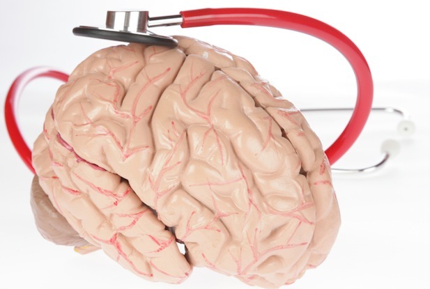
Diagnosing a Brain Injury

How do you diagnose a brain injury? There are a few different tests and assessments that can be administered to determine whether or not an individual may have sustained a brain injury. These tests and assessments include the Glasgow Coma Scale, radiological imaging and a neuropsychological assessment.
The Glasgow Coma Scale (“GCS”) is a 15 point test that measures a person’s ability to function and follow directions. The test looks specifically at the injured person’s coherence of speech and ability to move their eyes and limbs. A score of 3 means a person is unconscious. A lower score indicates a more severe brain injury. If a person’s GCS is below 9 this is likely a severe brain injury. A score between 9 and 12 usually indicates a moderate brain injury and a score of 13 and higher usually indicates a mild brain injury. This test is typically given by health care professionals to assess the initial severity of a brain injury. A GCS test is usually carried out immediately following the trauma (or as close to the event as possible).
There are also a variety of imaging technologies that can help identify a brain injury visually. One of these technologies is a computerized axial tomography scan (a.k.a CT or CAT scan) which uses x-rays to produce a series of images that show cross-sections of the brain. A CT scan can detect physical changes in the brain, such as hematomas (blood clots), hemorrhage (bleeding in the brain), contusions (bruised brain tissue) and swelling. However, CT scans often appear normal in patients with a mild traumatic brain injury.
Another imaging technology is magnetic resonance imaging (a.k.a MRI) test which uses magnets and radio waves to generate computerized images of the brain. MRI is more sensitive than CT scans but will still often appear normal in patients with a mild TBI.
A third type of imaging technology is the Single-photon emission computed tomography, (aka SPECT), which is a type of nuclear imaging and involves the injection of radioactive particles into the blood. Once the particles are injected a special camera rotates around the person and takes pictures from many angles. A computer uses these pictures to form a cross-sectional image. These cross sections can then be combined to create a 3D image of the brain and the blood flow to it. It has been said that a SPECT scan might be more sensitive to showing a brain injury than an MRI or CT because it can detect reduced blood flow to an injured site.
A neuropsychological examination is another method that can assist in diagnosing a brain injury, specifically a mild to moderate brain injury which may not have been detected using the imaging technologies mentioned above. This assessment involves many tests that are given and scored by a psychometrist and interpreted by a neuropsychologist. This assessment looks at an individual’s cognitive functioning while also taking into account their psychological state and personal circumstances.
If you suspect that you or a loved one has suffered a brain injury, it is important to consult a physician. If that injury happened due to the fault or neglect of someone else it is important to consult an experienced personal injury lawyer.
Contributors

A born-and-raised Barrie resident, Karen knows and loves her community. She is proud to be a partner in one of Canada’s most successful personal injury law firms—right in her own backyard. Karen joined Oatley Vigmond in 2013 as an associate lawyer. She holds a BA from Queen’s University and her Juris Doctor from Bond University in Australia. Prior to being called to the Bar in January 2013, Karen articled at a well-known personal injury law firm in Toronto.
