Making The Invisible Injury Visible In The Courtroom – Demonstrative Aids In Traumatic Brain Injury Cases
Why Demonstrative Evidence?
It is true that none of the strategies which I suggest is essential for success. Indeed you may be so convincing through the use of the spoken word that you have no need of demonstrative evidence. However, there is no denying that demonstrative evidence is especially and sometimes uniquely persuasive, that it is remembered and that it involves the listener. It may be that because many of the concepts a judge or juror must understand in a brain injury case are complex, that demonstrative evidence is particularly persuasive and therefore a strategy none of us can afford to overlook.
What is the Overall Objective?
The overall objective in a traumatic brain injury case is to convince the trier that an organic injury to the brain has occurred, that it is a permanent injury and that it is the main reason for your client’s impairment. In most cases proof of an organic injury is necessary in order to defeat one or all of the following standard defenses:
- Any impairment flows from pre-morbid, long standing emotional problems.
- Any impairment flows from some other event which occurred after the collision.
- If there was in injury, it was trivial, and any ongoing impairment is the result of an emotional reaction which can be easily cured.
- The plaintiff is malingering.
The Ambulance Record
If the ambulance record contains any evidence which is consistent with traumatic brain injury, it presents an opportunity for effective demonstrative evidence. The ambulance record can be converted to a transparency and projected onto a screen by an overhead projector for all to see. Witnesses who review the ambulance call report may highlight a copy of the record, which highlighted copy should be entered as an exhibit. The highlighted portion can be highlighted on the overhead transparency for emphasis in the minds of the juror.
You will wish to draw attention to any recording of a period of unconscious, any reference to disorientation as to person, place or time, and any reference to aggressive or agitated behavior. You will also wish to draw attention to any record of bruising or lacerations about the head. Finally, you will emphasize visually any Glasgow Coma Scale that supports your case.
The Emergency Record
The emergency records will include the initial history, a nurses’ history and assessment, a doctors’ history and assessment and a working diagnoses. If any references are made to disorientation, unconsciousness, dizziness, aggressive or agitated behavior, or other signs and symptoms of traumatic brain injury you will want to draw them to the trier’s attention by highlighting them on an overhead and on a copy of the record.
Remember that you may use the document itself as a piece of demonstrative evidence by creating a separate copy, having the witness highlight it in yellow and showing it to the jury after it is marked. Highlighting on the transparency copy projected onto the screen helps to reinforce through repetition.
If there is a significant Glasgow Coma Scale, you will, needless to say, highlight that as well and point out visually the comparison with the ambulance record.
Demonstrate Consistency by Reviewing Hospital Records and Clinical Notes
Demonstrative evidence techniques offer a marvelous technique for persuasively and efficiently reviewing nurses’ notes, doctors’ clinical notes and other records which have been compiled over a period of years. In this way you can show the trier of fact that the plaintiff’s presentation has been consistent and continuous since the collision. This consistency of presentation is logically incompatible, for most lay people, with emotional etiology and lends support to an organic diagnosis.
If, for example, a nurse notes right-sided headaches in hospital and the family doctor and neurologist made notes of similar complaints on and off for 3 years the conclusion becomes inescapable that there is an organic basis. Use of the overhead projector to show the jury or a judge the clinical notes, page by page, is the best way to present this evidence.
Each time that any of your experts refer to these early records take advantage of the opportunity to show them by way of the overhead projector to the trier – over and over again.
External Injury to the Head
Not surprisingly judges and jurors alike find it easier to believe the brain is injured if the head was injured. Not infrequently there are very early photos available which record black eyes, and cuts to the head. These photographs are important corroborative evidence which are especially effective when enlarged or when projected by use of an Elmo Presenter.
Later, when an expert is explaining the area of the brain which has been affected, it is persuasive to refer to the photograph and have the expert confirm that the effected part of the brain is adjacent to some laceration or bruising to the head.
Vehicle Damage
We all know how hard it is to persuade a judge or jury of physical injury when there is no visible damage to the plaintiff’s vehicle. The reverse is equally true, however. Whenever your client’s vehicle or the defendant’s vehicle is obviously damaged, photographs of the vehicle damage are powerful demonstrative evidence. If photographs are not available, project transparencies of the repair invoices to show the extent of the collision damage and collision forces.
Any obvious damage to the interior of the car caused by your client’s head should be demonstrated visually and discussed by every witness who can do so. Your client can draw attention to it as can any expert who is discussing the forces which impacted your client’s head.
When There is No Vehicle Damage
If there is no vehicle damage you must call a biomechanical engineer, supported by a reconstruction engineer’s estimate of speeds, to explain how the forces of this collision can cause brain injury. Demonstrative evidence is very effective as a device for explaining the nature of the forces and the impact of those forces on the brain within the skull.
There are any number of ways to create this evidence. Expensive and sophisticated computer animations are available, but tough to get into evidence. Medical-legal illustrations are effective. Many texts and articles contain simple stick drawings which are quite sufficient for explaining these concepts. The method I prefer, if your witness can draw, is to have him or her create the illustration on the Elmo Presenter as the oral evidence is given. By creating the evidence in front of the judge or juror the listener is involved and the evidence is therefore especially persuasive.
In Every Case, Explain the Mechanism of Injury
If you are to persuade the trier of fact that an injury has occurred you must find a way to overcome the appearance of normalcy as the plaintiff gives evidence. One way to do so is to graphically explain the mechanism of injury so that the jury or judge can appreciate that the brain can be permanently injured without any permanent change to the appearance of the head or face. There is no more effective way to explain the mechanism of injury that to show it, either through the use of medical-legal illustrations, animations or other artwork. Animations are best presented on videotaped. Illustrations can be projected from small originals or presented as mounted enlargements.
Evidence of Pre-Morbid Success
There is nothing more effective in persuading any trier of fact of organic injury than evidence that demonstrates that the plaintiff was having a great life. It follows as night the day that if the juror would be happy living the plaintiff’s life he or she will doubt that the plaintiff would choose to give it all up.
There are two powerful ways to use demonstrative evidence to create a visual image of a successful life. One way is through treatment, medication and employment charts which reveal graphically an absence of medical treatment and consistent employment before the injury.
Another way is through the use of a photographic essay of the plaintiff’s pre-morbid life. Nothing is more dramatic than photographs which portray the plaintiff as healthy, athletic, and prosperous. Family photographs or videos of special occasions, of teams, and of awards presentations are indisputable evidence with which every juror will identify. Photographs of the plaintiff’s home, cottage and other trappings of obvious success are effective. Equally dramatic are photographs of works or art or crafts which the plaintiff has created with his or her own hands. We enlarge these photographs so everyone in the courtroom can see the photograph with ease. I like to use them over an over again when I am examining lay witnesses and I find it effective to mount them on a stand beside the witness as a reminder of the image throughout the testimony. Sometimes if you use these photographs during cross-examination of defense experts they will help the juror recall all the evidence they heard from the plaintiffs’ lay evidence.
Comparison of Post-Traumatic Presentation
Photographs of your client three, four or five years after a brain injury will almost always offer a dramatic comparison. While is not proof of organic injury by itself, where the comparison is vivid the trier will ask how this change could occur without an organic basis. Enlarged comparative portraits mounted sided by side on a wafer board are the best way to present this evidence.
Use the Radiographic Evidence that is Available
There is a natural tendency to want to hide radiographic evidence which fails to demonstrate abnormality. This is a mistake, if only because the defence will make it seem as though it has found something which you attempted to hide.
I like to show the so-called “normal findings” in all their radiographic glory, either on the overhead projector or on Elmo. By doing so I demonstrate that the plaintiff has nothing to hide. Showing the radiograph presents an excellent opportunity for the expert to explain that the physician ordered the examination because of a belief that a traumatic brain injury had occurred. It also presents an excellent opportunity to explain the limitations of current radiographic techniques and to explain the functioning of the part of the brain that is the subject of the study.
Diagrams Which Simplify and Explain Radiographic Limitations
Since the defence will make the best of radiographs which are “normal” you must explain how an organic injury can exist in the absence of any finding. The best way to do so is by demonstrating visually how these imaging techniques work, because through an understanding of the technique comes an understanding of its limitations. Remember that to most laypersons an x-ray reveals all – and that the more sophisticated imaging like the CT or SPECT scan is infallible.
For example, the best way I know to take the sting out of a so-called “normal” CT scan is to show the juror a simple diagram of the slices through the brain which illustrate the layers of images used by this technique. The visual image of these slices, like the image of a slice of bread, is a powerful way to diffuse such a CT scan. Such an image is easily portrayed by use of an overhead projector or the Elmo Presenter.
An extremely simple way to make your point is to show the jury an x-ray of the head and to ask your expert if there is anyway to tell from the x-ray whether the patient has a headache.
Proof of the Limitations of Medical Science
Ignorance is your greatest enemy in these trials. As advocates we must remember that many judges and almost all jurors will be learning about brain injury for the first time in your trial. Most judges and jurors have far too much faith in the ability of doctors to diagnose and understand brain injury. One very effective way to explain how primitive is our knowledge is the use of brain function illustrations which were in common medical texts not long ago. Such an illustration enables an expert to explain to the trier of fact that we are still learning and still discovering things about the brain, on virtually a daily basis. This perspective is essential if the jury is to understand that a certain amount of faith is required and if the jury is to appreciate that medical knowledge in this field is not infallible.
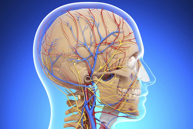
Functional Maps and Models
Another very powerful strategy in these cases rests on the theory that we can only believe in what we understand. There is no better way to assist a judge or juror to understand a brain injury than to provide a visual demonstration of how the brain functions. This demonstration is possible with the use of medical-legal illustrations or models of the brain.
The phenomenon of anchoring figures prominently in the use of demonstrative evidence. Your objective must be to establish a link between the functional impairment and the visual image of the affected part of the brain. Maps and models create this link if the link is reinforced in a consistent way throughout the trial.
It is especially persuasive to establish a link between the functional impairment, the area of the brain which must therefore be affected, and any visible injury to the head.
Explain Neuropsychological Tests and Limitations
Neuropsychological tests are simple if properly explained. However, left to a defense expert they may confound a judge or jury, or even worse, be used to create an impression that they support for the defense theory.
Demonstrative evidence can be used to unravel, simplify and communicate the findings of neuropsychological tests. Furthermore, by using a visual approach you have an opportunity to emphasize findings which support the conclusion of your expert that post – traumatic deficits exists.
The only limit to any demonstrative evidence is your own imagination. You might begin by projecting a transparency listing the tests employed in your neuropsychologists battery. The next transparency might compare the defense battery of tests. Transparencies can be used to illustrate visually the findings of each test and to compare the findings on the same tests by defense experts. For example, you might have a transparency which lists the results of consistency testing from each of the tests in the battery, which would provide a powerful demonstrative foundation for the conclusion that the plaintiff presented honestly and is not malingering.
By the time we get to trial it is not uncommon for the plaintiff to have seen several neuropsychologists and to have had multiple assessments. Without a chart for the jury to see a comparison of the results is virtually impossible. More importantly, an expert can get away with saying just about anything if the results are not properly understood.
Brain injuries almost always have devastating effects on their victims. To recover fair compensation it is essential to establish the organic basis of disability. Occasionally this can be challenging with even the severe brain injury because radiographic imaging is still so limited insofar as the brain is concerned. It is often challenging with moderate injuries and almost always with so-called mild brain injuries, where radiographic techniques cannot demonstrate injuries which are sometimes microscopic. Although challenging, you can meet the challenge of making the invisible become visible through the skillful use of demonstrative evidence. It is well worth the effort.